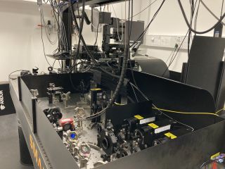
Total Internal Reflection Fluorescence Structured Illumination Microscope (TIRF-SIM)
eTIRF-SIM (extended Total Internal Reflection Fluorescence Structured Illumination) is a state of the art super-resolution microscope. It takes advantage of combining two imaging modalities TIRF and SIM in the same system, which allows achieving up to 90 nm axial and 150 nm lateral resolutions at very high acquisition rates – up to 100 ms per colour per frame. Moreover, TIRF illumination helps to reduce phototoxicity and photodamage as the sample is only illuminated at the basal plain leaving majority of it intact. This makes TIRF-SIM an ideal candidate for high resolution live cell imaging.
Physical changes, such as cytoskeleton rearrangements or protein/receptor localisations in the plasma membrane of cells, predominantly act on length-scales below the diffraction-limit of visible light. Conventional confocal microscopy therefore cannot resolve these processes and other super-resolution technology such as STED, even though they provide higher spatial resolutions, struggle to track these processes over long periods of time. The TIRF-SIM's ability to resolve these processes at the right spatial scales and monitor the underlying driving molecular rearrangements at the right temporal scales, are most obviously essential for mechanistic studies.
We combine TIRF-SIM with a toolbox of fluorescence microscopy techniques to investigate (i) healthy and pathological cell migration patterns, (ii) the detection of and interaction between immune cells, (iii) the 3D-molecular organisation and dynamics of immune relevant receptors essential for successful immune responses, and (iv) the dynamics of the primary cilium in health and disease such as rheumatoid arthritis. These experiments are just the starting point for comprehensive quantitative studies at high spatial-temporal resolutions in the context of health and disease.
MICROSCOPE OBJECTIVES:
- Olympus UApoN100× NA 1.49
- high NA 1.7 silicone immersion
ILLUMINATION:
- Lasers: 488 nm, 560 nm, and 650 nm
DETECTORS:
- 2 x Hamamatsu ORCA-Flash 4.0 v3 sCMOS
LIVE-CELL ENVIORMENT CONTROL:
- OKO lab incubator with temperature and CO2 control
SOFTWARE:
- Custom-built
ACKNOWLEDGMENT:
The TIRF-SIM microscope was built in collaboration with Micron, an Oxford-wide advanced microscopy technology consortium supported by Wellcome Strategic Awards (091911 and 107457) and an MRC/EPSRC/BBSRC next-generation imaging award. We also acknowledge Wellcome Trust (212343/Z/18/Z), and EPSRC (EP/S004459/1) grants.


